



- Home
- About
About
- Academics
acdemics
Faculties
-
Faculty of Arts and Commerce
- Department of Bengali
- Department of Commerce
- Department of English
- Department of Economics
- Department of Folklore
- Department of Hindi
- Department of History
- Department of Library & Information Science
- Department of Modern Language
- Department of Philosophy
- Department of Political Science
- Department of Rural Development Studies
- Department of Sanskrit
- Department of Sociology
- Department of Visual Arts
- Faculty of Science
- Faculty of Engineering Technology & Management
- Faculty of Education
- School of Interdisciplinary Studies
Academic Calendar
-
Faculty of Arts and Commerce
- Admission
Admission
- Administration
- Facilities
- Affiliated Colleges
Affiliated Colleges
- Centres
Centres
- Central Library
Central Library
- IQAC
- DODL
- Faculty
Faculty


Confocal Microscope
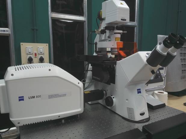
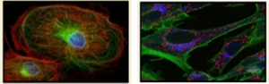
Confocal microscopes enable the fluorescence, bright field, and phase-contrast imaging of thick tissue samples. Light emitted from the sample either from illumination or laser stimulation of tagged dyes is filtered through a pinhole. This allows the focus of desired planes on the Z-stack as the rays emitted from different planes are blocked by the pinhole, eliminating out-of-focus glare. High-quality images can be obtained from specimens compared to conventional fluorescence microscopy and fixed and live samples can be imaged in real-time as well. The application includes research fields like stem cell research, fluorescence resonance energy transfer, fluorescence recovery after photo-bleaching, fluorescence in-situ hybridization, live cell imaging, co-localization studies, epitope tagging, etc.
NB: Minimum booking time is 1(One) hour
| Instrument | Usage Charge (INR) | |
| For Internal users | For External users | |
| General Imaging | Rs. 300 per hour | Rs. 600 per hour |
| Live Cell Imaging | Rs. 400 per hour Rs. 1000 for 4 hours |
Rs. 1500 per hour Rs. 2000 for 4 hours |